Canalicular Lacerations
Overview
- Canalicular lacerations are breaks (interruptions) in the normal tear duct drainage system. If not repaired promptly, tearing will usually lead to.
- This systems originates with the puncta (there is one in both the upper and the lower eyelid) and is a conduit for tears to travel from the eyelid through the nasolacrimal sac into the nose.
- Tension, from trauma such as a blow from the fist, can lead to in an eyelid laceration which involves the canalicular system.
- Repair requires re-approximation of the eyelid as well as re-approximation of the conduit; this is best achieved with a stent such as with silastic and fine sutures such as 6,7, or 8-0 vicryls.
.![Image of lacrimal trauma showing [description of objects] with embedded text that reads '[embedded text]'](/../topics/105-Lacrimal%20Trauma/Image/Capture.JPG)
1. Lacrimal Gland
2. Tear Film on the eye
3. Canalicular and Nasolacraiml duct
The photos below show a patient who was hit in their right eye with a fist and who sustained a canalicular laceration:
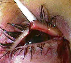
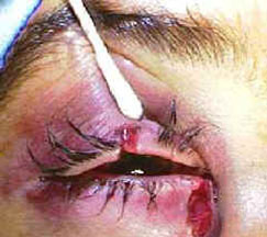
Treatment
There are several different means to repair such an injury. Placement of a stent (silastic tubing) helps maintain proper alignment of the conduit and prevent stricture after the repair.
- Bi-canalicular stent
This places places a silicone stent in both the traumatized (lacerated) canalicular system as well as the normal. One disadvantage of this technique is the potential damage to the "good" canalicular system.